
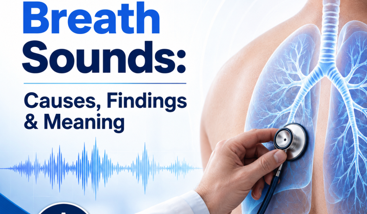
Bronchial Breath Sounds are a type of lung sound heard during a physical exam with a stethoscope. They are usually louder and harsher than normal breath sounds and are often heard over the upper chest or trachea. When they are heard in places where they normally should not be, they can suggest a lung problem such as pneumonia, collapse of lung tissue, or fluid-filled airspaces.
Understanding what bronchial breath sounds mean can help explain why a healthcare provider may order more tests or recommend treatment. This article explains the sound, what causes it, what it may indicate, and when it matters.
Table of Contents
- What Are Bronchial Breath Sounds?
- What Do They Mean?
- Common Causes
- What Do They Sound Like?
- Bronchial Breath Sounds vs Normal Breath Sounds
- How Doctors Interpret Them
- Other Physical Exam Findings
- Are They Dangerous?
- When to Seek Medical Attention
- How They Are Diagnosed
- Treatment Depends on the Cause
- In Children and Adults
- Key Takeaway
What Are Bronchial Breath Sounds?
Bronchial Breath Sounds are one of the normal breath sound types heard over the large airways, especially over the trachea and larynx. They are:
- Loud and high-pitched
- Harsh or hollow in quality
- Longer during exhalation than inhalation
- Usually heard best over the upper chest and neck
In a healthy lung exam, these sounds are not typically heard over the peripheral lung fields. Instead, doctors usually hear vesicular breath sounds in most areas of the lungs, which are softer and gentler.
What Do They Mean?
The meaning depends on where they are heard.
Normal finding
If bronchial breath sounds are heard over the trachea or main airways, this is normal.
Abnormal finding
If they are heard over the lung tissue, especially near the chest wall, they may indicate that air is moving through lung tissue that has become more solid than normal. This can happen when the air sacs are filled with fluid, pus, or tissue, or when lung tissue is collapsed but the airway remains open.
In simple terms, bronchial breath sounds in the wrong location may mean the lungs are not air-filled in the usual way.
Common Causes of Bronchial Breath Sounds
Bronchial Breath Sounds over the lungs are often associated with conditions that increase the density of lung tissue. Common causes include:
1. Pneumonia
Pneumonia is one of the most common causes. In bacterial pneumonia, part of the lung can fill with inflammatory fluid, making breath sounds sound more bronchial.
Other findings may include:
- Fever
- Cough
- Shortness of breath
- Crackles or egophony
- Chest pain
2. Lung Consolidation
Consolidation means lung tissue has become more solid, usually because air spaces are filled with fluid, blood, pus, or cells. Pneumonia is a frequent cause, but consolidation can also occur with other lung diseases.
3. Atelectasis
Atelectasis is collapse of part of the lung. Breath sounds may be heard near the affected area, especially if the airways remain open while the surrounding lung tissue collapses.
4. Pulmonary Fibrosis or Other Lung Scarring
Scarring can change how sound travels through the lungs. This is less common than pneumonia, but abnormal breath sounds may be present in affected areas.
5. Lung Mass or Tumor
A mass can alter airflow and sound transmission. Depending on the location and size, bronchial breath sounds may be noted over the affected region.
6. Pleural Effusion with Adjacent Consolidation
Fluid around the lung usually reduces breath sounds, but if there is nearby collapsed or consolidated lung tissue, bronchial sounds may still be heard in adjacent areas.
For a broader overview of related respiratory findings, see this overview of normal and abnormal lung sounds from the NCBI Bookshelf.
What Do They Sound Like?
People often ask what bronchial breath sounds are like on auscultation. They are described as:
- Harsh
- Tubular
- Hollow
- High-pitched
- Loud
They are usually more noticeable during exhalation than inhalation. This is the opposite of normal vesicular breath sounds, which are softer and more prominent during inhalation.
Bronchial Breath Sounds vs Normal Breath Sounds
A quick comparison helps make the difference clear.
Vesicular breath sounds
- Soft and low-pitched
- Heard over most of the lungs
- Inhalation is longer than exhalation
- Normal finding in peripheral lung fields
Bronchial Breath Sounds
- Loud and high-pitched
- Heard normally over the trachea
- Exhalation is as long as or longer than inhalation
- Abnormal if heard over peripheral lung tissue
Bronchovesicular breath sounds
These are a middle type:
- Moderate pitch and intensity
- Normally heard over the main bronchi and between the shoulder blades
- Equal inspiratory and expiratory phases
Understanding these differences helps explain why the location of the sound is so important.
How Doctors Interpret Bronchial Breath Sounds
A healthcare provider does not interpret this finding alone. They look at it together with:
- The patient’s symptoms
- Temperature and oxygen level
- Cough or sputum production
- Chest imaging, such as a chest X-ray
- Other lung sounds, such as crackles or wheezing
For example, a harsh breath sound with fever and cough may strongly suggest pneumonia. Abnormal sounds after surgery or prolonged bed rest may point toward atelectasis.
Clinicians often compare the exam with chest imaging, and the brain damage after heart attack article is one example of how timely evaluation can matter when oxygen levels are affected, even though the underlying condition is different.
Other Physical Exam Findings That May Occur
They may be found alongside other signs, such as:
- Crackles: often from fluid or inflammation in the lungs
- Egophony: when the spoken “E” sounds like “A”
- Bronchophony: louder, clearer voice transmission through consolidated lung
- Whispered pectoriloquy: whispered words sound unusually distinct
- Dullness to percussion: may suggest consolidation or pleural fluid
- Reduced chest expansion: may occur with lung collapse or severe infection
These findings help confirm whether the sound reflects consolidation, collapse, or another issue.
Are Bronchial Breath Sounds Dangerous?
They are not dangerous by themselves. They are a physical exam finding, not a disease. However, they may point to an underlying condition that needs treatment.
They are more concerning when:
- Heard in peripheral lung areas
- Associated with shortness of breath
- Accompanied by fever or chest pain
- Found in someone with low oxygen levels
- Present after surgery or prolonged immobility
The importance of the finding depends on the full clinical picture.
When to Seek Medical Attention
You should get medical care if they are suspected along with symptoms such as:
- Trouble breathing
- Chest pain
- High fever
- Persistent cough
- Coughing up mucus or blood
- Blue lips or fingertips
- Confusion
- Worsening fatigue
These symptoms may indicate pneumonia, lung collapse, or another serious lung condition.
How They Are Diagnosed
These breath sounds are identified during a chest exam with a stethoscope. A doctor or clinician listens to both sides of the chest and compares the sound patterns.
If abnormal sounds are found, further evaluation may include:
- Chest X-ray
- CT scan
- Blood tests
- Pulse oximetry
- Sputum testing
- Additional respiratory assessment
The exam finding is only one part of the diagnosis.
Treatment Depends on the Underlying Cause
There is no specific treatment for the sound itself. The goal is to treat what is causing it.
If pneumonia is the cause
Treatment may include:
- Antibiotics, if bacterial
- Rest and fluids
- Fever control
- Oxygen if needed
If atelectasis is the cause
Treatment may include:
- Deep breathing exercises
- Incentive spirometry
- Early movement
- Treating pain that limits breathing
- Addressing mucus blockage
If another lung condition is present
Care depends on the diagnosis and may include medications, oxygen therapy, or further testing.
Early treatment matters for lung conditions that reduce oxygen exchange, which is why clinicians also watch for changes that can affect the lungs in serious systemic illness, such as brain stem stroke warning signs, recovery and treatment.
Bronchial Breath Sounds in Children and Adults
They are interpreted the same way in children and adults: location matters most. In both groups, hearing these sounds over the outer lung fields is usually abnormal.
In children, abnormal breath sounds may appear more quickly or become more noticeable because smaller airways can be affected by infection or mucus buildup more easily.
Key Takeaway
Bronchial Breath Sounds are normally heard over the trachea and major airways, but if they are heard over the lungs, they often suggest an abnormality such as pneumonia, consolidation, or atelectasis. The finding is important because it helps doctors identify where lung tissue may be filled with fluid, collapsed, or otherwise changed.
If they are noted along with breathing difficulty, fever, chest pain, or low oxygen, medical evaluation is important. The sound itself is not the diagnosis, but it can be an important clue to an underlying lung condition.
More on bronchial breath sounds and lung exam findings
When clinicians talk about bronchial breath sounds, they are usually describing a specific pattern heard during auscultation rather than a separate disease. That distinction matters because the same sound can be normal in one area of the body and abnormal in another.
Over the trachea, the sound is expected because the air is moving through large central airways. Over the lung periphery, however, the same sound suggests that the normal air-filled pattern of the lung has changed. In many cases, this happens when inflammation or fluid reduces the amount of air in the alveoli.
That is why the examiner does not rely on the sound alone. The physical exam is always interpreted together with the history, including whether the person has a sudden or gradual onset of symptoms, recent infection, surgery, chest trauma, or prolonged immobility. Even a simple change in breathing pattern can help a clinician narrow the cause.
Why location matters
The same breath sound can mean very different things depending on where it is heard. This is one of the most important ideas in respiratory examination. A sound that is expected over the neck may be a red flag over the lower lung zones.
In practical terms, the clinician maps what is heard across the chest. If bronchial quality is limited to the central airways, it is usually normal. If it is heard in a focal area of the lungs, it often points to a localized process such as consolidation or collapse. If it is heard more widely, the clinician may consider a larger area of infection, scarring, or another structural change.
This approach helps explain why lung auscultation remains useful even in the age of imaging. The stethoscope cannot replace a chest X-ray or CT scan, but it can quickly suggest where to look first.
Pneumonia and consolidation
Pneumonia is a classic reason for bronchial breath sounds to appear over the lung fields. When infection causes the alveoli to fill with inflammatory material, sound travels differently through that area. The result may be a louder, harsher, more tubular quality on examination.
Consolidation is a related concept. It describes lung tissue that has become denser than normal, regardless of the exact cause. Although pneumonia is the most common cause, consolidation can also follow bleeding into the lung, inflammatory disease, or other processes that replace air with material inside the alveoli.
Because the sound can be localized, a clinician may detect bronchial breath sounds over one lobe while the rest of the lung exam remains relatively normal. That pattern can be useful when deciding whether imaging is needed.
Atelectasis and airway blockage
Atelectasis often develops when a portion of the lung collapses or does not fully expand. This can happen after surgery, with shallow breathing due to pain, or when mucus blocks an airway. In these situations, the surrounding tissue may transmit sound in an unusual way.
If the airway remains open, the sound may still travel through the region and create a bronchial quality. This is one reason atelectasis can sometimes resemble pneumonia on exam, especially when symptoms are limited or early.
In practice, other clues help separate the causes. Fever and productive cough raise the likelihood of infection, while recent surgery, immobility, or poor inspiratory effort may point more toward collapse.
What doctors look for next
After hearing abnormal breath sounds, clinicians often check for a few additional details:
- Is the sound localized or widespread?
- Does it change after coughing or deep breathing?
- Are there signs of airway obstruction or mucus retention?
- Is oxygen saturation reduced?
- Are there symptoms outside the lungs, such as fever, chills, or fatigue?
These details help determine whether the person needs urgent care, antibiotics, breathing support, or further imaging. In many cases, the breath sound is the first clue rather than the final answer.
Recovery and prevention
There is no direct way to prevent bronchial breath sounds because they are a finding, not a condition. Prevention depends on avoiding the illnesses that cause them when possible.
General steps that support lung health include:
- Seeking early care for respiratory infections
- Following post-surgical breathing instructions
- Getting up and moving after prolonged bed rest when safe to do so
- Managing chronic lung disease as directed
- Avoiding smoking and secondhand smoke
Recovery also depends on the cause. Someone with simple atelectasis may improve with deep breathing and movement, while a person with pneumonia may need antibiotics and close follow-up. If the sound reflects a more serious lung or heart-related problem, treatment may need to be more intensive.
Bottom line
Bronchial Breath Sounds are normal over the trachea and major airways, but when they are heard over the lung fields, they can signal a problem such as pneumonia, consolidation, or atelectasis. The key question is not just what the sound is, but where it is heard and what other findings are present.
When combined with symptoms like fever, cough, chest pain, or shortness of breath, the finding deserves prompt medical attention. By itself, it is not a diagnosis, but it can be an important sign that helps guide the next step in care.